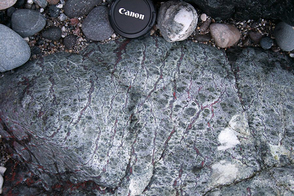
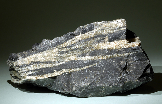
Serpentinite is a metamorphic rock that is mostly composed of serpentine group minerals. Serpentine group minerals antigorite, lizardite, and chrysotile are produced by the hydrous alteration of ultramafic rocks.
serpentinization (is a geological low-temperature metamorphic process ) involves chemical reactions which convert unhydrous ferromagnesian silicate minerals (pyroxene, olivine) into hydrous silicate minerals (serpentine) plus some other possibilities like brucite and magnetite. Brucite forms if the precursor rocks are rich in magnesium (dunite, for example). Magnetite forms if there is enough iron present (pyroxenite). Usually serpentinite contains iron in the form of magnetite which gives dark color to serpentinites.
Serpentinization is accompanied by release of heat (it is an exothermic reaction); the heat released by a complete Serpentinization reaction of 1 Kg of peridotites is 250 Joules, enough to bring the temperature of 1 Liter of Water up by 50°C degrees under normal temperature and pressure conditions. Serpentinization is also accompanied by a change in the physical properties of the rock; the volume increase by up 30 % with concomitant decrease in density (a non Serpentinized peridotites has a density of 3300Kg/m
.
In parallel, the speed of propagation of seismic waves decrease during Serpentinization: from nearly 8Km/s in non serpentinized rocks, to 5.5Km/s in serpentinites. The production of magnetite during Serpentinization is accompanied by a increase of the rock’s magnetic susceptibility; finally, serpentine is less tolerant to deformation than olivine and pyroxenes; 15-20% of serpentine in peridotites is enough to decrease the rock’s tolerance to deformation and can specifically facilitate the motion on the faults.
Chrysotile Asbestos Veins. From Natalie Teager, Arizona State University.
.jpg)
Serpentinite from Italian Alps. From www.sandatlas.org
.jpg)
Serpentinite from Canada. From www.sandatlas.org

Serpentinized Peridotite , Lizard complex, Cornwall. From Department of Earth Sciences (Oxford).
• David Shelley (1983): Igneous and metamorphic rocks under the microscope. Campman & Hall editori.
• E. WM. Heinrich (1956): Microscopic Petrografy. Mcgraw-hill book company,inc
• Anthony R. Phillpotts & Jay J. Ague: Principles of igneous and metamorphic petrology. Cambridge editore.
• Passchier, Cees W., Trouw, Rudolph A. J: Microtectonics (2005)
Photo
.jpg)
Relict Olivine (High relief) surrounded by (pale green-yellow) in a Serpentinite. PPL image, 2x (Field of view = 7mm) |
.jpg)
Relict Olivine (High relief) surrounded by (pale green-yellow) in a Serpentinite. PPL image, 2x (Field of view = 7mm) |
.jpg)
Relict Olivine (High relief) surrounded by (pale green-yellow) in a Serpentinite. PPL image, 2x (Field of view = 7mm) |
.jpg)
Relict Olivine (High relief) surrounded by (pale green-yellow) in a Serpentinite. PPL image, 10x (Field of view = 2mm) |
.jpg)
Relict Olivine (High relief) surrounded by (pale green-yellow) in a Serpentinite. PPL image, 10x (Field of view = 2mm) |
.jpg)
Relict Olivine (High relief) surrounded by (pale green-yellow) in a Serpentinite. PPL image, 10x (Field of view = 2mm) |
.jpg)
Green Spinel in a Serpentinite. PPL image, 2x (Field of view = 7mm) |
.jpg)
Green Spinel in a Serpentinite. PPL image, 2x (Field of view = 7mm) |
.jpg)
Green Spinel in a Serpentinite. PPL image, 2x (Field of view = 7mm) |
.jpg)
Green Spinel in a Serpentinite. PPL image, 2x (Field of view = 7mm) |
.jpg)
Green Spinel in a Serpentinite. PPL image, 10x (Field of view = 2mm) |
.jpg)
Green Spinel in a Serpentinite. PPL image, 10x (Field of view = 2mm) |
.jpg)
Olivine with Mesh Structure in a Serpentinized Peridotite. PPL image, 2x (Field of view = 7mm) |
.jpg)
Olivine with Mesh Structure in a Serpentinized Peridotite. XPL image, 2x (Field of view = 7mm) |
.jpg)
Olivine with Mesh Structure in a Serpentinized Peridotite. PPL image, 2x (Field of view = 7mm) |
.jpg)
Olivine with Mesh Structure in a Serpentinized Peridotite. XPL image, 2x (Field of view = 7mm) |
.jpg)
Olivine with Mesh Structure in a Serpentinized Peridotite. PPL image, 10x (Field of view = 2mm) |
.jpg)
Olivine with Mesh Structure in a Serpentinized Peridotite. XPL image, 10x (Field of view = 2mm) |
.jpg)
Olivine with Mesh Structure in a Serpentinized Peridotite. PPL image, 2x (Field of view = 7mm) |
.jpg)
Olivine with Mesh Structure in a Serpentinized Peridotite. XPL image, 10x (Field of view = 2mm) |
.jpg)
Olivine with Mesh Structure in a Serpentinized Peridotite. PPL image, 10x (Field of view = 2mm) |
.jpg)
Olivine with Mesh Structure in a Serpentinized Peridotite. XPL image, 10x (Field of view = 2mm) |
.jpg)
Completely serpentinized Olivine. PPL image, 10x (Field of view = 2mm) |
.jpg)
Completely serpentinized Olivine. XPL image, 10x (Field of view = 2mm) |
.jpg)
Olivine with Mesh Structure in a Serpentinized Peridotite. XPL image, 10x (Field of view = 2mm) |
.jpg)
Olivine with Mesh Structure in a Serpentinized Peridotite. XPL image, 10x (Field of view = 2mm) |
.jpg)
Olivine with Mesh Structure in a Serpentinized Peridotite. PPL image, 10x (Field of view = 2mm) |
.jpg)
Olivine with Mesh Structure in a Serpentinized Peridotite. XPL image, 10x (Field of view = 2mm) |
.jpg)
Olivine with Mesh Structure in a Serpentinized Peridotite. XPL image, 10x (Field of view = 2mm) |
.jpg)
Olivine with Mesh Structure in a Serpentinized Peridotite. XPL image, 10x (Field of view = 2mm) |
.jpg)
<
Olivine with Mesh Structure in a Serpentinized Peridotite. XPL image, 10x (Field of view = 2mm) |

Olivine with Mesh Structure in a Serpentinized Peridotite. XPL image, 10x (Field of view = 2mm) |
.jpg)
Olivine with Mesh Structure in a Serpentinized Peridotite. XPL image, 10x (Field of view = 2mm) |
.jpg)
Olivine with Mesh Structure and brown pyroxene in a Serpentinized Peridotite. PPL image, 2x (Field of view = 7mm) |
.jpg)
Olivine with Mesh Structure in a Serpentinized Peridotite. XPL image, 10x (Field of view = 2mm) |
.jpg)
Olivine with Mesh Structure and brown pyroxene in a Serpentinized Peridotite. PPL image, 2x (Field of view = 7mm) |
.jpg)
Olivine with Mesh Structure and brown pyroxene in a Serpentinized Peridotite. XPL image, 2x (Field of view = 7mm) |
.jpg)
Olivine with Mesh Structure and brown spinel in a Serpentinized Peridotite. PPL image, 10x (Field of view = 2mm) |
.jpg)
Olivine with Mesh Structure and brown spinel in a Serpentinized Peridotite. PPL image, 10x (Field of view = 2mm) |
.jpg)
Olivine with Mesh Structure in a Serpentinized Peridotite. XPL image, 10x (Field of view = 2mm) |
.jpg)
Olivine with Mesh Structure in a Serpentinized Peridotite. XPL image, 10x (Field of view = 2mm) |
.jpg)
Olivine with Mesh Structure in a Serpentinized Peridotite. XPL image, 10x (Field of view = 2mm) |
.jpg)
Deformed Pyroxene in a Tectonized peridotite. XPL image, 2x (Field of view = 7mm) |
.jpg)
Deformed Pyroxene in a Tectonized peridotite. PPL image, 2x (Field of view = 7mm) |
.jpg)
Deformed Pyroxene in a Tectonized peridotite. XPL image, 2x (Field of view = 7mm) |
.jpg)
Deformed Pyroxene in a Tectonized peridotite. XPL image, 2x (Field of view = 7mm) |
.jpg)
Deformed Pyroxene in a Tectonized peridotite. PPL image, 2x (Field of view = 7mm) |
.jpg)
Deformed Pyroxene in a Tectonized peridotite. XPL image, 2x (Field of view = 7mm) |
.jpg)
Pyroxene (pale beige) altered Olivine (pale yellow) and fibrous, colorless amphibole crystas (secondary amphibole). PPL image, 2x (Field of view = 7mm) |
.jpg)
Pyroxene (pale beige) altered Olivine (pale yellow) and fibrous, colorless amphibole crystas (secondary amphibole). PPL image, 2x (Field of view = 7mm) |
.jpg)
Pyroxene (pale beige) altered Olivine (pale yellow) and fibrous, colorless amphibole crystas (secondary amphibole). PPL image, 2x (Field of view = 7mm) |
.jpg)
Sermpentine veins in a serpentinite from Elba island. XPL image, 2x (Field of view = 7mm) |
.jpg)
Sermpentine veins in a serpentinite from Elba island. XPL image, 2x (Field of view = 7mm) |
.jpg)
Olivine with Mesh Structure in a Serpentinized Peridotite. PPL image, 2x (Field of view = 7mm) |
.jpg)
Olivine with Mesh Structure in a Serpentinized Peridotite. PPL image, 2x (Field of view = 7mm) |
.jpg)
Olivine with Mesh Structure in a Serpentinized Peridotite. PPL image, 2x (Field of view = 7mm) |
.jpg)
Olivine with Mesh Structure in a Serpentinized Peridotite. XPL image, 2x (Field of view = 7mm) |
.jpg)
Olivine with Mesh Structure and pale beige pyroxene in a Serpentinized Peridotite. PPL image, 2x (Field of view = 7mm) |
.jpg)
Olivine with Mesh Structure and pyroxene in a Serpentinized Peridotite. XPL image, 2x (Field of view = 7mm) |
.jpg)
Olivine with Mesh Structure and pale beige pyroxene in a Serpentinized Peridotite. PPL image, 2x (Field of view = 7mm) |
.jpg)
Rounded olivine (completaly altered by serpentine) and brown altered pyroxene. note the radial fractures, due to the increase in volume during the alteration of olivine in serpentine. PPL image, 2x (Field of view = 7mm) |
.jpg)
Rounded olivine (completaly altered by serpentine) and altered pyroxene. XPL image, 2x (Field of view = 7mm) |
.jpg)
Rounded olivine (completaly altered by serpentine) and brown altered pyroxene. note the radial fractures, due to the increase in volume during the alteration of olivine in serpentine.PPL image, 2x (Field of view = 7mm) |
.jpg)
Rounded olivine (completaly altered by serpentine) and brown altered pyroxene. XPL image, 2x (Field of view = 7mm) |
.jpg)
Rounded olivine (completaly altered by serpentine) and brown altered pyroxene. note the radial fractures, due to the increase in volume during the alteration of olivine in serpentine. PPL image, 2x (Field of view = 7mm) |
.jpg)
Rounded olivine (completaly altered by serpentine) and altered pyroxene. XPL image, 2x (Field of view = 7mm) |
.jpg)
Rounded olivine (completaly altered by serpentine) and brown altered pyroxene. note the radial fractures, due to the increase in volume during the alteration of olivine in serpentine. PPL image, 2x (Field of view = 7mm) |
.jpg)
Rounded olivine (completaly altered by serpentine) and altered pyroxene. XPL image, 2x (Field of view = 7mm) |
.jpg)
Rounded olivine (completaly altered by serpentine) and brown altered pyroxene. note the radial fractures, due to the increase in volume during the alteration of olivine in serpentine. PPL image, 2x (Field of view = 7mm) |
.jpg)
Serpentinite from Heidelberg in Baden (Germany). XPL image, 2x (Field of view = 7mm) |
.jpg)
Serpentinite from Heidelberg in Baden (Germany). XPL image, 2x (Field of view = 7mm) |
.jpg)
Serpentinite from Heidelberg in Baden (Germany). XPL image, 2x (Field of view = 7mm) |
.jpg)
Serpentinite from Heidelberg in Baden (Germany). XPL image, 2x (Field of view = 7mm) |
.jpg)
Serpentinite from Heidelberg in Baden (Germany). XPL image, 2x (Field of view = 7mm) |
.jpg)
Serpentinite from Heidelberg in Baden (Germany). XPL image, 2x (Field of view = 7mm) |
.jpg)
Serpentinite from Heidelberg in Baden (Germany). XPL image, 2x (Field of view = 7mm) |
.jpg)
Serpentinite from Heidelberg in Baden (Germany). XPL image, 2x (Field of view = 7mm) |
.jpg)
Serpentinite from Heidelberg in Baden (Germany). XPL image, 2x (Field of view = 7mm) |
.jpg)
Serpentinite from Heidelberg in Baden (Germany). XPL image, 2x (Field of view = 7mm) |
.jpg)
Serpentinite from Heidelberg in Baden (Germany). XPL image, 2x (Field of view = 7mm) |
.jpg)
Serpentinite from Heidelberg in Baden (Germany). XPL image, 10x (Field of view = 2mm) |
.jpg)
Serpentinite. Elba Island. XPL image, 2x (Field of view = 7mm) |
.jpg)
Serpentinite. Elba Island. XPL image, 10x (Field of view = 2mm) |
.jpg)
Serpentinite. Elba Island. XPL image, 10x (Field of view = 2mm) |
.jpg)
Serpentinite. Elba Island. XPL image, 2x (Field of view = 7mm) |
.jpg)
Serpentinite. Elba Island. XPL image, 2x (Field of view = 7mm) |
.jpg)
Serpentinite. Elba Island. XPL image, 10x (Field of view = 2mm) |
.jpg)
Serpentinite. Elba Island. PPL image, 2x (Field of view = 7mm) |
.jpg)
Serpentinite. Elba Island. PPL image, 2x (Field of view = 7mm) |
.jpg)
Serpentinite. Elba Island. XPL image, 2x (Field of view = 7mm) |
.jpg)
Serpentinite. Elba Island. PPL image, 2x (Field of view = 7mm) |
.jpg)
Serpentinite. Elba Island. PPL image, 2x (Field of view = 7mm) |
.jpg)
Serpentinite. Livorno (Tuscany), Italy. PPL image, 2x (Field of view = 7mm) |
.jpg)
Serpentinite. Livorno (Tuscany), Italy. PPL image, 2x (Field of view = 7mm) |
.jpg)
Serpentinite. Livorno (Tuscany), Italy. PPL image, 2x (Field of view = 7mm) |
.jpg)
Serpentinite. Livorno (Tuscany), Italy. PPL image, 2x (Field of view = 7mm) |
.jpg)
Sermpentine veins in a serpentinite from Livorno (Tuscany), Italy. PPL image, 2x (Field of view = 7mm) |
.jpg)
Sermpentine veins in a serpentinite from Livorno (Tuscany), Italy. XPL image, 2x (Field of view = 7mm) |
.jpg)
Sermpentine veins in a serpentinite from Livorno (Tuscany), Italy. PPL image, 2x (Field of view = 7mm) |
.jpg)
Sermpentine veins in a serpentinite from Livorno (Tuscany), Italy. XPL image, 2x (Field of view = 7mm) |
.jpg)
Sermpentine veins in a serpentinite from Livorno (Tuscany), Italy. XPL image, 2x (Field of view = 7mm) |
.jpg)
Sermpentine veins in a serpentinite from Livorno (Tuscany), Italy. XPL image, 2x (Field of view = 7mm) |
.jpg)
Sermpentine veins in a serpentinite from Livorno (Tuscany), Italy. XPL image, 2x (Field of view = 7mm) |
.jpg)
particular of sermpentine veins in a serpentinite from Livorno (Tuscany), Italy. XPL image, 10x (Field of view = 2mm) |
.jpg)
particular of sermpentine veins in a serpentinite from Livorno (Tuscany), Italy. XPL image, 10x (Field of view = 2mm) |
.jpg)
Serpentinized pyroxene in a serpentinite from Livorno (Tuscany), Italy. XPL image, 10x (Field of view = 2mm) |
.jpg)
Serpentinized pyroxene in a serpentinite from Livorno (Tuscany), Italy. XPL image, 10x (Field of view = 2mm) |
.jpg)
Sermpentine veins in a serpentinite from Livorno (Tuscany), Italy. XPL image, 2x (Field of view = 7mm) |
.jpg)
Sermpentine veins in a serpentinite from Livorno (Tuscany), Italy. XPL image, 2x (Field of view = 7mm) |
.jpg)
Sermpentine veins in a serpentinite from Livorno (Tuscany), Italy. XPL image, 10x (Field of view = 2mm) |
.jpg)
Sermpentine veins in a serpentinite from Livorno (Tuscany), Italy. XPL image, 2x (Field of view = 7mm) |
.jpg)
Cavity filld by carbonates in a serpentinite from Livorno (Tuscany), Italy. PPL image, 2x (Field of view = 7mm) |
.jpg)
Cavity filld by carbonates in a serpentinite from Livorno (Tuscany), Italy. XPL image, 2x (Field of view = 7mm) |
.jpg)
Cavity filld by carbonates in a serpentinite from Livorno (Tuscany), Italy. PPL image, 2x (Field of view = 7mm) |
.jpg)
Rounded olivine (completaly altered by serpentine) and brown altered pyroxene. note the radial fractures, due to the increase in volume during the alteration of olivine in serpentine. Champdepraz, Valle d'Aosta, Italy. PPL image, 2x (Field of view = 7mm) |
.jpg)
Rounded olivine (completaly altered by serpentine) and brown altered pyroxene . Champdepraz, Valle d'Aosta, Italy. XPL image, 2x (Field of view = 7mm) |
.jpg)
Rounded olivine (completaly altered by serpentine) and brown altered pyroxene. note the radial fractures, due to the increase in volume during the alteration of olivine in serpentine. Champdepraz, Valle d'Aosta, Italy. PPL image, 2x (Field of view = 7mm) |
.jpg)
Rounded olivine (completaly altered by serpentine) and brown altered pyroxene. note the radial fractures, due to the increase in volume during the alteration of olivine in serpentine. Champdepraz, Valle d'Aosta, Italy. PPL image, 2x (Field of view = 7mm) |
.jpg)
Rounded olivine (completaly altered by serpentine) and brown altered pyroxene . Champdepraz, Valle d'Aosta, Italy. XPL image, 2x (Field of view = 7mm) |
.jpg)
Rounded olivine (completaly altered by serpentine) and brown altered pyroxene. note the radial fractures, due to the increase in volume during the alteration of olivine in serpentine. Champdepraz, Valle d'Aosta, Italy. PPL image, 2x (Field of view = 7mm) |

.jpg)
.jpg)


.jpg)
.jpg)
.jpg)
.jpg)
.jpg)
.jpg)
.jpg)
.jpg)
.jpg)
.jpg)
.jpg)
.jpg)
.jpg)
.jpg)
.jpg)
.jpg)
.jpg)
.jpg)
.jpg)
.jpg)
.jpg)
.jpg)
.jpg)
.jpg)
.jpg)
.jpg)
.jpg)
.jpg)
.jpg)
.jpg)
.jpg)

.jpg)
.jpg)
.jpg)
.jpg)
.jpg)
.jpg)
.jpg)
.jpg)
.jpg)
.jpg)
.jpg)
.jpg)
.jpg)
.jpg)
.jpg)
.jpg)
.jpg)
.jpg)
.jpg)
.jpg)
.jpg)
.jpg)
.jpg)
.jpg)
.jpg)
.jpg)
.jpg)
.jpg)
.jpg)
.jpg)
.jpg)
.jpg)
.jpg)
.jpg)
.jpg)
.jpg)
.jpg)
.jpg)
.jpg)
.jpg)
.jpg)
.jpg)
.jpg)
.jpg)
.jpg)
.jpg)
.jpg)
.jpg)
.jpg)
.jpg)
.jpg)
.jpg)
.jpg)
.jpg)
.jpg)
.jpg)
.jpg)
.jpg)
.jpg)
.jpg)
.jpg)
.jpg)
.jpg)
.jpg)
.jpg)
.jpg)
.jpg)
.jpg)
.jpg)
.jpg)
.jpg)
.jpg)
.jpg)
.jpg)
.jpg)
.jpg)
.jpg)
.jpg)
.jpg)
.jpg)
.jpg)
.jpg)
.jpg)
.jpg)
.jpg)
.jpg)