Boudinage
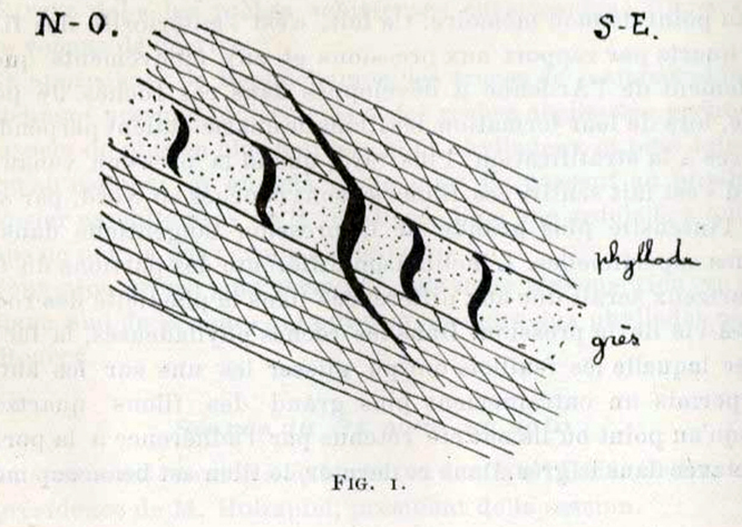
The term boudins, which comes from the French word for sausage, was first used in 1908 by Max Lohest for shortened boudins or mullions in Bastogne in Belgium. Before that, boudins and pinch-and-swell structures had been described several times. The exact definition and meaning of the terms boudins and boudinage have changed through much of the twentieth century, but there is now consensus that boudins are extensional structures formed by layer-parallel extension, while boudinage is the process that leads to the formation of boudins from originally continuous layers.

An illustration of the original "boudins" in the report of the field meeting in Bastogne of the Société Géologique de Belgique (Lohest et al. 1908).
Classic boudins form where single competent layers are extended into separate pieces through plastic, brittle or a combination of plastic and brittle deformation mechanisms (
Fig.1). The boudinaged layer is located in a rock matrix that deforms plastically. Boudins are separated by brittle extension fractures (left side) or by shear fractures that may be symmetric or asymmetric (middle and right side of
Fig.1). Instead of fractures, boudins may also be separated by narrow ductile shear zones that are confined to the boudinaged layer.
.png)
Fig.1: The geometry of boudins is largely controlled by whether boudins are separated by extension or shear fractures, and the influence of plastic versus brittle deformation mechanisms. Asymmetric boudins may indicate non coaxial deformation. From Structural Geology, Fossen, H. (2010)
There are also examples of regularly spaced areas of thinning in many extended competent layers without the separation into isolated fragments or boudins. Such structures are called pinch-and-swell structures and the process by which they form is known as necking. Pinch-and-swell structures are structures where the boudin-like elements are barely connected, as shown in
Fig.2. Both regular boudins and pinch-and-swell structures are controlled by temperature, strain rate and viscosity contrast or a well-developed foliation. High temperature promotes plastic deformation mechanisms also in the most competent layer. High-viscosity contrast and strain rate promote fracturing of competent layers. These parameters will also affect the geometry of boudins.

Fig.2: Descriptional terminology of boudins (a) and pinch-and-swell structures (b). From Structural Geology, Fossen, H. (2010)
Boudins come in different shapes (
Fig.3), depending on how they behaved during deformation. The different shapes relate to the competence contrast across the boudinaged layer and its two side, and of the type of fracture that forms (extension or shear fractures). Rectangular boudins form when the boudins do not deform internally, when the boudinaged layer behaves completely stiff or brittlely. Barrel-shaped boudins and fish-mouth boudins arise when the boudins deform plastically along their margins. Unicorn boudins form when the competence contrast is higher on one side of the boudinaged layer, and lower on the other side (stress is concentrated in the corner regions of boudins, that’s why the corners tend to deform more easily than the rest of the boudins).
.png)
Fig.3: Different types of boudin geometry, all involving extensional fracturing; (a) rectangular, (b) barrel-shaped, (c) fish-mouth, (d) unicorn. From Structural Geology, Fossen, H. (2010)
Bibliography
• Bucher, K., & Grapes, R. (2011). Petrogenesis of metamorphic rocks. Springer Science & Business Media.
• Fossen, H. (2016). Structural geology. Cambridge University Press.
• Howie, R. A., Zussman, J., & Deer, W. (1992). An introduction to the rock-forming minerals (p. 696). Longman.
• Passchier, Cees W., Trouw, Rudolph A. J: Microtectonics (2005).
• Philpotts, A., & Ague, J. (2009). Principles of igneous and metamorphic petrology. Cambridge University Press.
• Shelley, D. (1993). Igneous and metamorphic rocks under the microscope: classification, textures, microstructures and mineral preferred-orientations.
• Vernon, R. H. & Clarke, G. L. (2008): Principles of Metamorphic Petrology. Cambridge University Press.
• Vernon, R. H. (2018). A practical guide to rock microstructure. Cambridge university press.
Photo

Boudinaged pelitic layer in a marble. XPL image, 10x (Field of view = 2mm) |
(1).jpg)
Boudinaged calcite vein. Elba island, Italy. PPL image, 10x (Field of view = 2mm) |
(8).jpg)
Boudinaged calcite vein. Elba island, Italy. PPL image, 10x (Field of view = 2mm) |
(5).jpg)
Boudinaged calcite vein. Notice how matrix flows around the boudins. Elba island, Italy. PPL image, 2x (Field of view = 7mm) |
(6).jpg)
Boudinaged calcite vein. Notice how matrix flows around the boudins. Elba island, Italy. PPL image, 2x (Field of view = 7mm) |
(2).jpg)
Boudinaged calcite vein. Notice how matrix flows around the boudins. Elba island, Italy. PPL image, 2x (Field of view = 7mm) |
(3).jpg)
Boudinaged calcite vein. Notice how matrix flows around the boudins. Elba island, Italy. PPL image, 2x (Field of view = 7mm) |
(4).jpg)
Boudinaged calcite vein. Notice how matrix flows around the boudins. Elba island, Italy. PPL image, 2x (Field of view = 7mm) |
(7).jpg)
Boudinaged calcite vein. Notice how matrix flows around the boudins. Elba island, Italy. PPL image, 2x (Field of view = 7mm) |
.jpg)
Boudinaged plagioclase-rich layer in amphibolite. Italian Alps. PPL image, 1x (Field of view = 9mm) |
.jpg)
Boudinaged plagioclase-rich layer in amphibolite. Italian Alps. PPL image, 1x (Field of view = 9mm) |
.jpg)
Boudinaged plagioclase-rich layer in amphibolite. Italian Alps. PPL image, 2x (Field of view = 7mm) |
.jpg)
Boudinaged plagioclase-rich layer in amphibolite. Italian Alps. PPL image, 2x (Field of view = 7mm) |
.jpg)
Boudinaged plagioclase-rich layer in amphibolite. Italian Alps. PPL image, 2x (Field of view = 7mm) |
.jpg)
Boudinaged plagioclase-rich layer in amphibolite. Italian Alps. PPL image, 10x (Field of view = 2mm) |
.jpg)
Boudinaged amphibole crystal. Merano, Tyrol, Italian Alps. PPL image, 2x (Field of view = 7mm) |
.jpg)
Boudinaged amphibole crystal. Merano, Tyrol, Italian Alps. PPL image, 2x (Field of view = 7mm) |
.jpg)
Boudinaged amphibole crystal. Merano, Tyrol, Italian Alps. XPL image, 2x (Field of view = 7mm) |
.jpg)
Boudinaged amphibole crystal. Merano, Tyrol, Italian Alps. PPL image, 2x (Field of view = 7mm) |
.jpg)
Boudinaged amphibole crystal. Merano, Tyrol, Italian Alps. XPL image, 2x (Field of view = 7mm) |
.jpg)
Boudinaged amphibole crystal. Merano, Tyrol, Italian Alps. PPL image, 2x (Field of view = 7mm) |
.jpg)
Boudinaged amphibole crystal. Merano, Tyrol, Italian Alps. XPL image, 2x (Field of view = 7mm) |
.jpg)
Boudinaged amphibole crystal. Merano, Tyrol, Italian Alps. PPL image, 2x (Field of view = 7mm) |
.jpg)
Boudinaged amphibole crystal. Merano, Tyrol, Italian Alps. XPL image, 2x (Field of view = 7mm) |
.jpg)
Boudinaged amphibole crystal. Merano, Tyrol, Italian Alps. PPL image, 10x (Field of view = 2mm) |
.jpg)
Boudinaged amphibole crystal. Merano, Tyrol, Italian Alps. PPL image, 10x (Field of view = 2mm) |
.jpg)
Boudinaged amphibole crystal. Merano, Tyrol, Italian Alps. XPL image, 10x (Field of view = 2mm) |
.jpg)
Boudinaged amphibole crystal. Merano, Tyrol, Italian Alps. PPL image, 10x (Field of view = 2mm) |
.jpg)
Boudinaged amphibole crystal. Merano, Tyrol, Italian Alps. XPL image, 10x (Field of view = 2mm) |
.jpg)
Boudinaged amphibole crystal. Merano, Tyrol, Italian Alps. PPL image, 10x (Field of view = 2mm) |
.jpg)
Boudinaged amphibole crystal. Merano, Tyrol, Italian Alps. XPL image, 10x (Field of view = 2mm) |
.jpg)
Boudinaged amphibole crystal. Merano, Tyrol, Italian Alps. PPL image, 10x (Field of view = 2mm) |
.jpg)
Boudinaged amphibole crystal. Merano, Tyrol, Italian Alps. PPL image, 10x (Field of view = 2mm) |
.jpg)
Boudinaged amphibole crystal. Merano, Tyrol, Italian Alps. XPL image, 10x (Field of view = 2mm) |
.jpg)
Boudinaged amphibole crystal. Merano, Tyrol, Italian Alps. PPL image, 10x (Field of view = 2mm) |
.jpg)
Boudinaged amphibole crystals. Merano, Tyrol, Italian Alps. PPL image, 10x (Field of view = 2mm) |
.jpg)
Boudinaged amphibole crystals. Merano, Tyrol, Italian Alps. XPL image, 10x (Field of view = 2mm) |
.jpg)
Boudinaged amphibole crystal. Merano, Tyrol, Italian Alps. PPL image, 10x (Field of view = 2mm) |
.jpg)
Boudinaged amphibole crystal. Merano, Tyrol, Italian Alps. XPL image, 10x (Field of view = 2mm) |
.jpg)
Boudinaged amphibole crystal. Merano, Tyrol, Italian Alps. PPL image, 10x (Field of view = 2mm) |
.jpg)
Boudinaged amphibole crystal. Merano, Tyrol, Italian Alps. XPL image, 10x (Field of view = 2mm) |
.jpg)
Boudinaged amphibole crystal. Between boudin neck grows fibrous amphibole. Merano, Tyrol, Italian Alps. PPL image, 20x (Field of view = 1mm) |
.jpg)
Boudinaged amphibole crystal. Between boudin neck grows fibrous amphibole. Merano, Tyrol, Italian Alps. XPL image, 20x (Field of view = 1mm) |
.jpg)
Boudinaged amphibole crystal. Between boudin neck grows fibrous amphibole. Merano, Tyrol, Italian Alps. PPL image, 20x (Field of view = 1mm) |
.jpg)
Boudinaged amphibole crystal. Between boudin neck grows fibrous amphibole. Merano, Tyrol, Italian Alps. XPL image, 20x (Field of view = 1mm) |
.jpg)
Big boudinaged allanite crystal. Note the deflection of the muscovite layer near the boudin neck. PPL image, 10x (Field of view = 2mm) |
.jpg)
Big boudinaged allanite crystal. Note the deflection of the muscovite layer near the boudin neck. PPL image, 2x (Field of view = 7mm) |
.jpg)
Big boudinaged allanite crystal. Note the deflection of the muscovite layer near the boudin neck. PPL image, 2x (Field of view = 7mm) |
.jpg)
Big boudinaged allanite crystal. Note the deflection of the muscovite layer near the boudin neck. XPL image, 2x (Field of view = 7mm) |
.jpg)
Big boudinaged allanite crystal. Note the deflection of the muscovite layer near the boudin neck. PPL image, 2x (Field of view = 7mm) |
.jpg)
Big boudinaged allanite crystal. Note the deflection of the muscovite layer near the boudin neck. XPL image, 2x (Field of view = 7mm) |
.jpg)
Big boudinaged allanite crystal. Note the deflection of the muscovite layer near the boudin neck. PPL image, 2x (Field of view = 7mm) |
.jpg)
Big boudinaged allanite crystal. Note the deflection of the muscovite layer near the boudin neck. XPL image, 2x (Field of view = 7mm) |
.jpg)
Big boudinaged allanite crystal. Note the deflection of the muscovite layer near the boudin neck. PPL image, 2x (Field of view = 7mm) |
.jpg)
Big boudinaged allanite crystal. Note the deflection of the muscovite layer near the boudin neck. XPL image, 2x (Field of view = 7mm) |

.png)

.png)


(1).jpg)
(8).jpg)
(5).jpg)
(6).jpg)
(2).jpg)
(3).jpg)
(4).jpg)
(7).jpg)
.jpg)
.jpg)
.jpg)
.jpg)
.jpg)
.jpg)
.jpg)
.jpg)
.jpg)
.jpg)
.jpg)
.jpg)
.jpg)
.jpg)
.jpg)
.jpg)
.jpg)
.jpg)
.jpg)
.jpg)
.jpg)
.jpg)
.jpg)
.jpg)
.jpg)
.jpg)
.jpg)
.jpg)
.jpg)
.jpg)
.jpg)
.jpg)
.jpg)
.jpg)
.jpg)
.jpg)
.jpg)
.jpg)
.jpg)
.jpg)
.jpg)
.jpg)
.jpg)
.jpg)
.jpg)
.jpg)